

Dr. Mitch Broser
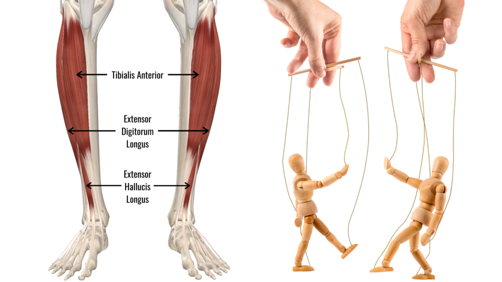
The “anterior compartment” of the lower leg includes the tibialis anterior, extensor digitorum long, and extensor hallucis longus. These muscles contribute to ankle dorsiflexion, but their unique locations and attachments produce variations in the movement.
The tibialis anterior is the bulky, tubular muscle that lives right beside your prominent shin bone. As this muscle runs down the leg, it takes a turn across the front of the ankle to the inside, where it wraps under your foot. The contraction of this muscle brings your foot up into dorsiflexion and rotates your foot into inversion.
The extensor digitorum longus lives just to the outside of the tibialis anterior. This muscle runs down the leg, then close to the ankle, it splits into 4 tendons where they attach into each of the lateral 4 toes. This muscle contracts to dorsiflex the ankle and extend the lateral 4 toes.
The extensor hallucis longus is the smallest of the three and is largely hidden deep, under the tibialis anterior and extensor digitorum longus. As it travels down the leg, it moves from deep to superficial between the tibialis anterior and extensor digitorum longus tendon and runs down to the end of the big toe. This muscle produces big toe dorsiflexion, or extension, along with ankle dorsiflexion.
Together, like strings on a marionette, these 3 muscles work together to control the movement of the ankle and foot into dorsiflexion. When these muscles get over-worked with repetitive movements and inadequate recovery, the tissue quality of the anterior leg compartment declines. Over time, the tissues that hold the muscle fibers together lose elasticity, lowering the amount of force they can handle, and in turn, lowering their tissue failure (injury) threshold.
Not only does it affect the function of the individual muscle-tendon unit – it also affects the stretching and loading of the neighboring muscles! Force can be transmitted from muscle to muscle, and with poor overloading, multiple muscles and tissue can become problematic. This can lead to poor relative movement between muscles. When a muscle can’t move freely from its neighbours, it will pull on the neighbouring muscles and tissues, causing inefficient muscle function and tissue overloading. Like a marionette with a few strings twisted together, this will limit independent control of the anterior leg compartment muscle, leading to dysfunction and inevitable injury (acute injury and progressive overload injury) if left unmanaged.
The muscles and tissues of the anterior leg commonly become problematic in runners, hikers, and avid walked. The human gait cycle demands for loading of the anterior leg compartment muscles and tissue through absorbing force with every step. Acute injuries and chronic dysfunctions of the leg can accelerate tissue overloading. The only way to improve tissue quality and strength of the anterior leg compartment is with tissue-specific training. By understanding the anatomy of the anterior leg and the mechanisms to alter tissue architecture for the better, we can enhance the force-absorption capacity and raise the injury threshold to mitigate your risk of a lower leg injury, and keep moving better.